Diagnostic Clinical Reasoning and treatment approach: An ExaFmple of how Evidence Practice and individualised treatment and Care is Approached. Upper Limb.

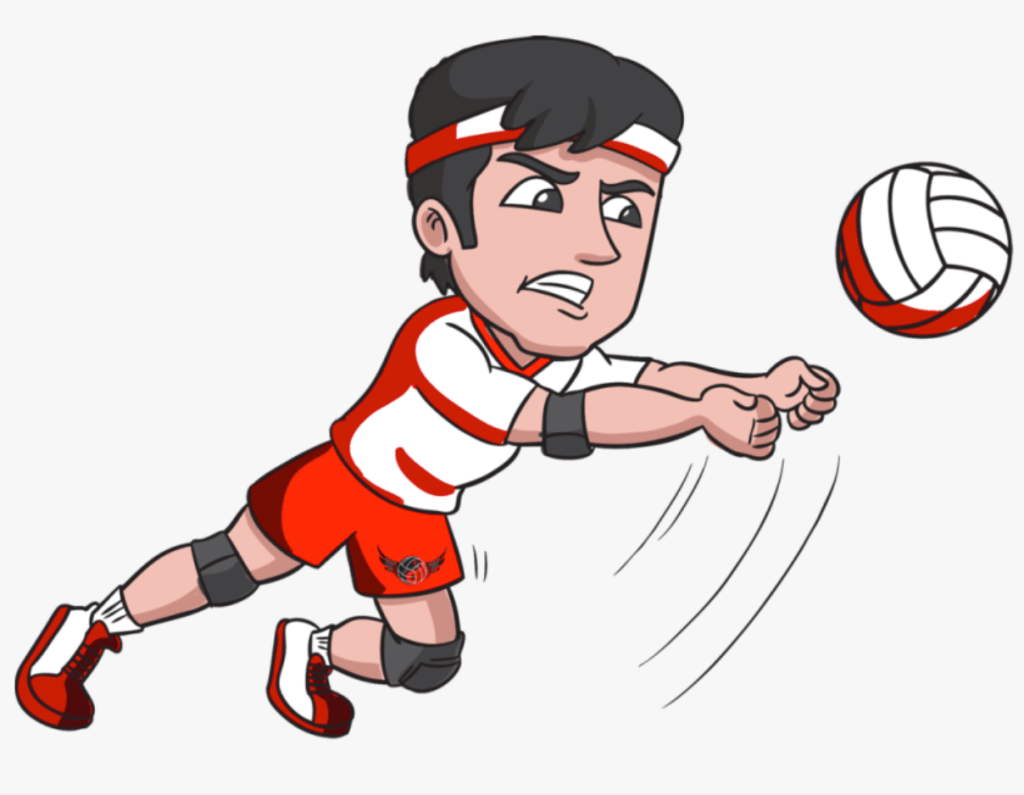
A 22-year-old elite female rugby player complains of acute pain after making a tackle with an outstretched arm. She comes off the pitch cradling her arm.

Anatomical and biomechanical features that contribute to stability of the shoulder.
The shoulder is a synovial joint. It features three bones. The Scapula, humerus and the clavicle. It compromises the Glenohumeral, Sternoclavicular, Acromioclavicular and Scapulothoracic joints. Shoulder stability is characterised by static and dynamic soft tissues. These structures work together to keep the head of the humerus within the glenoid. Instability can happen when the shoulder joint that hold the capsule together is stretched. (glenoid ligaments and/or glenoid labrum) Static stability is also achieved through the negative pressure in the shoulder joint creating a relative vacuum. (1)Muscle tone is also an important static stabiliser of the shoulder (2). Dynamic stabilisers work together to maintain the integrity of position, within the framework of the static stabilisers. Dynamic stabilisers emphasize the contractile and active movement of tendons and muscles.
Glenohumeral ligaments function as agents of stabilisation at the extreme ranges of motion, and become increasingly important as other structures are weakest (end range of musculature). There are three main ligaments in this joint. Superior ligament and middle ligament, limiting External rotation. Middle ligaments also helps limit anterior translation.
Inferior ligament being the thickest has an anterior and posterior band. Other ligaments include the coracohumeral ligament and coracromial ligament.
Dynamic contractile musculature includes the deltoid, scapular and rotator cuff muscles. They help facilitate a sensorimotor system that enacts efficient movement for neuromuscular stability. Dynamic stability is predicated on the shoulder maintain resistance to translation from opposite side muscle pull, support of muscles from muscle tensioning and synergistic force coupling. (3).
Muscles can be divided into Intrinsic (deeper) and extrinsic (superficial) muscles. Intrinsic include the supraspinatus, infraspinatus, subscapularis and Teres minor. Extrinsic muscles can include Latissimus dorsi, teres major Pectoralis major and minor. Secondary movers include the biceps and triceps. Primary dynamic stabilisers include the rotator-cuff muscles; Long head of biceps and Deltoid. Secondary stabilisers include Latissimus Dorsi, Teres Major and Pectoralis Major (4).
All these structures and muscles work together to maintain the Scapulohumeral rhythm, causing more refined movement, at a larger range of motion, and is characterised as a ratio of the glenohumeral to scapulothoracic movement to facilitate fluidity of motion. (5)
Shoulder Classification System:
There are several classification systems that have been used to historically classify shoulder stability. The Stanmore Triangle by Lewis et al. (6)identified three main groups. Types include: Polar I-Traumatic/Structural; Polar II-atraumatic Structural; Polar III – Muscle Patterning Non-structural. In figure-1 we can see the Stanmore classification showing a continuum between each group, with patients falling in between. (6)

(6)
The system accommodates for the way patients can fall in-between the poles. Has a graduation from traumatic to atraumatic, and takes into account muscle patterning and structural cases. Across each end of the triangle we have two subgroups. (6) The classification is important in order to correctly diagnose what type of instability the patient has and what management to utilise. Surgical reconstruction is recommended when structural damage is present, and the patient falls within the Polar-I group. The polar-III group recommendation is to avoid surgery and focus on muscle repatterning through physical rehabilitation. Polar-II is more controversial as it often shares an aspect of muscle repatterning.
Our patient is engaged in a contact sport and has Likely injured themselves acutely and traumatically with an outstretched arm into contact. The tackle was completed; however the amount of force is uncertain and whether the shoulder was vulnerable to begin with due to inefficient muscle patterning. In most cases however with a good history of injury, of a high-level rugby player, with acute injury, we would classify as a Polar-1 injury.(7)
Differential diagnoses and rasoning in this athlete as she is brought off the pitch
Chrichton et al. (2012) shows that in the ‘tackler’ Group with an outstretched abducted shoulder, the most common injuries were glenohumeral dislocation, labral injuries and rotator cuff tears. (8)
Diff. Diagnosis: Most likely it is a Dislocation of the shoulder (anterior most likely) will take athlete off the field. Possible avulsion fracture of the glenoid could be an aspect of the dislocation. Subluxation might also be the case but less painful (might not come off the field). Labrum tear may present with less pain, and might continue game. This may also be possible with a superior labrar tear from anterior to posterior (SLAP) lesion, which is an injury to the glenoid labrum.
Subjective and objective assessment findings that you would expect in an anterior dislocation and explain the radiological imaging findings that might confirm diagnosis.
Subjective assessment includes an expansion of patient history. This includes Past Medical History, this can also help rule out red flags. History of present acute condition is important (previous subluxation or other shoulder pathology – Recurring injury). This can help us classify our management options based on the Stanmore classification system.Feeling of looseness in abduction can be associated with looseness can indicate glenohumeral instability.
Mechanism of injury is very important too. Description of arm position, together with the type of impact force, and weather the athlete experienced pain in the tackle, or on the fall can help in the differential diagnosis too. Palpation can help indicate presence of swelling, temperature. Bony prominences can be palpated over “AC-SC Joints, Acromion, bicep tendon, acromion and greater tuberosity.” (9). Indication of asymmetry pain reproduction as well as differences in sensation. Objective assessment should consider an observation on symmetry. This includes shape, position and function. Provocative exam manoeuvres for detection of anterior dislocation can be seen through the identification of a sulcus sign test. Hyperabduction test can test integrity of IGHL. The anterior apprehension test can be used to test for guarding. The Relocation test can be positive when there is relief of guarding. Anterior release test again used to assess for guarding. The load and shift test can detect anterior laxity. (9)
Radiological imaging includes the x-ray to ascertain for any fracture. It is also used to check for dislocation, weather anterior or inferior. And an X-ray can confirm your findings.If there is a fracture CT scan is useful to identify the details of the fracture. If there are more serious concerns and surgery is indicated MRI is indicates and is used because it gives more details of soft tissue injuries and can help guide the surgeon. In conservative treatment is indicated, the x-ray is usually enough. However, it must be considered that since she is a young elite female rugby player, at a young age who is high risk for re-injury. And therefore, will likely benefit from an MRI because she will likely need surgery to avoid possibility of recurrent dislocations (10).
Anterior Dislocation confirmed, and the shoulder was successfully relocated. The player then had a surgical stabilization. Early, middle and late stage rehab for this player.
Early stage management (6 weeks) includes immobilisation in an internally rotated and adducted position. Protection of surgical repair and achievement of protected ROM and patient education. Important goals in the early phase is to decrease pain and inflammation. Strengthening and proprioception focus. Utilisation of isometric strengthening. Rehabilitation to be continued with scapular strength and neuromuscular control. External rotation to 30 degrees. Outcome measures to be achieved include a protected ROM, Reduction in paid/inflammation as well as adequate stability. (11)
In the middle stage (6-12 weeks) we want to increase proprioception as well as strengthen dynamic stabilisers. The focus should be an increase in muscular strength in rotator cuff, as well as progress scapular activation and strengthening. Neuromuscular activation focus. We need to work within our achieved ROM and utilise motion. Outcome measures for progression to next stage, includes minimal pain, symmetrical capsule mobility as well as full ROM without pain with exception of end-range 90 degree abduction. Improve weaknesses in dynamic stability of scapulothoracic muscles. Progress to overhead positions as well a complex-movements with progressions in both speed and load. In this stage we need to start generic fitness for the elite athlete as able. (12)
In the late stage we need to improve neuromuscular control with increased focus of advancement of strength and start training for power. Increase endurance and include plyometrics. Advance general fitness too, to not fall behind and be able to return to play sooner. In full contact sports like rugby we need to be able to facilitate impact-loading of the glenohumeral joint by improving metrics in strength, power and stability. These athletes may need more time than the general population of other sports to return to play of less demanding sports. (13)
Finally, we need to prepare the athlete for return to play, with sport specific drills and a sport specific program (12).
An 18 year-old volleyball player comes to see you 72 hours after falling onto the point of his shoulder. He complains of pain and swelling over the superior aspect of his shoulder and has been unable to sleep on his right side.
Functional anatomy of the acromioclavicular joint, and the factors hat contribute to stability of the ACJ.
The acromioclavicular joint is a diarthrodial joint. The two bones are connected by a capsule inside the joint. There is a meniscus (intra-articular disc). Stability of the joint is structurally held together by the congruency of the bones and ligaments. The coraco-acromial ligament is present as well the trapezoid and conoid ligament. These are all static stabilisers. The muscles help with dynamic stabilisation. Specifically, the deltoid is very important for the anterior clavicle and the trapezius for the acromion. The symbiotic relationship of all the above muscles, work together and contribute to shoulder stability.
The possible differential diagnoses in this scenario and explain the features of the history which might influence your reasoning.
The mechanism of injury as described in the history, (Direct injury) is the most common mechanism that can cause acromioclavicular (AC) joint injury, through subluxation or dislocation.
Indirect Injury on an outstretched hand or elbow is a lot rarer. Possible differential diagnosis includes Subluxation of the AC joint; Dislocation of the AC joint; Fracture of the end of the clavicle; Fracture of the acromion.
Rupturing the rotator cuff, although unlikely, must be excluded. The mechanism of falling on the point of his shoulder at the acromion together with presentations of pain and swelling over the superior aspect of the shoulder, makes the AC joint injury the most likely diagnosis.
Examination of the athlete and the findings that would help to distinguish between differential diagnoses above.
Careful examination is necessary as there can always be the possibility of a fracture. History of patient is important in the subjective assessment. Through the objective assessment we can observe through the eye and palpation any spelling in the front and superior aspect of the joint.
Before examining the shoulder, we should clear the cervical spine and elbow joint. If normal we can continue with our clinical examination and evaluate the glenohumeral joint. Observation of local swelling and possible deformity and/or abrasions may be present. Deformity in the skin (tent skin) may be observed if the displacement is severe enough. (14)
Palpation is used as tolerated by patient. The proximal end of the clavicle can be palpated to see if there is an abnormal movement in the distal end. Gentle palpation of the inflamed area can be used to check for an increased pain response or any crackling sounds.
If no signs of fracture, the clavicle could be reduced. By stabilising the proximal third of the clavicle with one hand and elevating the scapula by pushing under the elbow with the other hand, horizontal stability can be deduced.
Performing the Scarf test is not necessary in an acute injury due to pain and discomfort. Any of the above tests should be abandoned if there is too much pain and proceed with a radiological evaluation.
If injury is present only in the AC-joint, then the range of movement in the glenohumeral joint is possible as long as there is no displacement in the AC joint below 90 degrees abduction and flexion.
Classification system of ACJ injuries and explain the radiological imaging which could help determine the classification.
The Rockwood classification in 1998 (15) is the most popular system for acromioclavicular joint injuries. It is a 6-tier classification system which was modified from the earlier 3 tier system by Allman, 1972 (16) and Tossy systems, 1963 (17).
Toast/Allman Types I-III were described as follows:
“Type I= AC sprain
Type II = complete rupture AC and sprain CC with less than 100% displacement.
Type III= AC AND CC ruptured. Clavicle displaced 100% of its width”
(16)
Rokwood, that is more helpful to decide for the surgeon if surgery is necessary is as follows:
“I. Sprain AC ligament
II. complete rupture AC ligament
III. Complete rupture AC AND CC ligament
IV. As above plus posterior movement of the clavicle through trapezius
V. Complete rupture. Grossly displaced superiority PLUS rupture of attachments of deltoid and trapezius.
VI. Inferior sub-coracoid displacement.” (15)
Based on the Rockwood system, type 1 and II do not need surgery. Recommended treatment is often physiotherapy and sling Immobilisation. Type III injuries have more controversy. Conservative treatment is recommended, and if there is no improvement surgery should be considered. With regards to Type IV, V, VI a referral to an orthopaedic surgeon is necessary for surgical management.
We can classify based on radiological imaging, with the use of an x-ray. The Zanca view (15 degrees cephalic tilt); AC view and weight bearing imaging. Weight bearing can be done by asking the patient to hold 1kg of weight with the patient relaxed. Weight bearing imaging is best for identifying type I and II cases. (18)
The management options for this athlete and the factors which may influence management.
Type 1 and 2 usually should adhere to conservative treatment and the POLICE protocol. (Protection, Optimal Loading, Ice and Elevation.) A sling is desired to immobilise and elevate the arm.
Return to daily living activities for type 1 injuries is usually at 2-4 weeks. 4 to 6 weeks with regards to type 2 injuries. 6-12 weeks when injury is type 3. (19)
Rehabilitation protocols are varied and based on each patient tolerance. In the Acute phase, we should focus on ROM, moving from passive, to active-assisted and then active ROM exercises.
We will then progress to proximal stability, followed by strength in the sub-acute phase
Finally we can accommodate sport specific conditioning in the late stage.
Type 3 is controversial and different orthopaedic units follow different protocols of management especially in high profile athletes. Usually starts with physical therapy and evaluated in the process accordingly. (20)
Type 4,5,6 in Rockwood classification is usually surgical and he should be sent to an orthopaedic centre specialising in these injuries. (21)
After surgery should be given specific attention with physiotherapy to reach the full ROM and full strengthening of the shoulder girdle muscles before going back to action. We should accommodate for protection of the surgical repair, control pain, reduce swelling (cold packs- 15-20 min several times a day), work to achieve ROM (Removal of sling for pendulum passive). Protocol and management would progress from ROM to strength training and finally dynamic movements and perturbation type exercise.
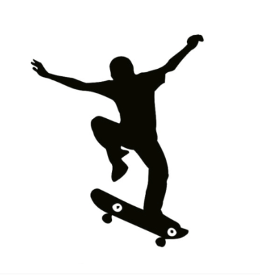
A 25 year-old skateboarder presents with a painful and stiff wrist 2 weeks after falling heavily on his wrist. He has continued training through the pain but has noticed recurrent swelling.
Possible differential diagnoses in this scenario and explain the features of the history which might influence your reasoning.
Possible differential diagnoses after falling heavily on the wrist but continuing training through the pain include a fractured scaphoid (unidsplaced); scapholunate dissociation; Lunotriqutral instability, Radiocarpal and midcarpal ligament injury (non-dissociative); Fracture of distal radius (non-displaced); Triangular fibrocartilage complex (TFCC) injury.
Mechanism of injury can vary, in position of wrist, and rotation.
Clinical assessment and investigations that would be appropriate to differentiate between differential diagnoses.
Clinical assessment of the wrist after such an injury involves inspection of the area looking for any deformity or swelling.
We should examine the wrist carefully checking for any clinical sign starting with the scaphoid.
We can utilise the ‘snuff box test’ looking for tenderness of the scaphoid and tenderness over the distal radius which could be fractured.
In a scapholunate ligament dissociation the bone of the proximal row do not move together – Scaphoid shift test (20% of normal people have a positive test). (22)
In radial deviation the scaphoid flexes and the lunate and triquetrum flex with it. (23)
We should also test for triangular fibrocartilage complex tear with a Compression test. We could examine – Finkelstein test for De Quervains pathology (unusual after an injury).
Useful investigation are plain x-rays which can inform on most pathologies including, scaphoid fracture, radial fractures. They can also show dynamic views of the wrist and can also show scapho-lunate dissociation.
Magnetic Resonance Imaging can indicate bone oedema and sometimes inter-carpal ruptured ligaments can be detected. Triangular fibrocartilage complex tear (TFL) can also at times be detected, despite it being inconsistent at times.
A wrist can also be symptomatic and radiographs of both x-ray and MRI negative. Arthroscopy may be indicated and is considered a gold standard of care, that can also help in for making an efficient management plan. (24)
The biomechanics of the proximal row of carpal bones during functional wrist motion and the effect of injury on these movements.
The bones in the proximal carpal row are the Scaphoid, lunate, Triquetrim and Pisiform. The proximal carpal row moves and adapts. It functions as a bridge between the forearm and the hand.
Rupture of the scapholunate ligament can cause Schapholunate dissociation, lunotriquetral dissociation or a radial column orientated force can cause fracture of the scaphoid. Both injuries disrupt the function of the proximal carpal row. (25).
Fracture of the scaphoid usually occurs in active young adults. If there is carpal instability, then the wrist is unable to keep its normal alignment and the destruction of intercarpal ligaments in the proximal row. This causes instability that affects the movement, power and function of the hand and wrist. (26)
The most likely fracture associated with this injury and the management options available. The advantages and disadvantages of these options.
The most likely fracture associated with this injury is a scaphoid fracture. The scaphoid fracture does not heal very well. It depends explicitly where the fracture occurred and has a direct dependency with the blood supply of the bone. One could elect to treat both Surgically or conservatively. A conservative non-surgical approach will avoid asscosiated complications of surgery, including anaesthesia, infection, discomfort, however it may need 3-4 months in a cast, with a possibility of non-union. Surgical treatment of the scaphoid with a special scaphoid screw gives the opportunity of early movement and increases the possibility of bone union. The associated advantages of early movement is an accelerated rehabilitation process and may be suitable for the athlete who wants a quicker return. Long term results, and complication rates seem similar. (27)
The athlete reports a previous history of a ‘jersey finger’ injury during five-a-side football. The pathophysiology and presentation of this injury.
Jersey finger may also be called ‘rugby finger’. It is an avulsion of the FDP – attached at the base of the distal phalanx. It usually happens when grabbing another players jersey with the tips of one or more fingers and the other player is in motion. Surgical treatment of jerseys finger is almost always required. It generally is indicative an 8-12 week period of no-contact sports.
In Presentation pain and tenderness presents at volar distal finger. Slight extension of injured finger is presented in relation to other fingers in a resting position. There is no active flexion of distal interphalangeal joint (DIP). Flexor tendon could be palpated to observe for retraction proximally through flexor sheath. (28)
The Classification system for Jersey finger is Leddy and Packer classification. Injuries range from 5 types. Type 1 would need surgery in the period of 7-10 days. Type 2-3 would need surgical repair within a few weeks for a desired outcome. Overall, surgery should be done within less than 3 weeks.(29)
If the injury is older than 3 months, a salvage procedure known as DIP arthrodesis could be attempted.
References:
1. Habermeyer P, Schuller U, Wiedemann E. The intra-articular pressure of the shoulder: An experimental study on the role of the glenoid labrum in stabilizing the joint. Arthroscopy. 1992;8(2):166-72.
2. VP K, P B. The role of atmospheric pressure in stabilising the shoulder. An experimental study. The Journal of Bone and Joint Surgery British volume. 1985;67-B(5):719-21.
3. Biel A, Dorn R. Trail guide to the body: How to locate muscles, bones and more: Andrew Biel; 1997.
4. Itoi E, Newman SR, Kuechle DK, Morrey BF, An K. Dynamic anterior stabilisers of the shoulder with the arm in abduction. The Journal of bone and joint surgery British volume. 1994;76(5):834-6.
5. Struyf F, Nijs J, Baeyens JP, Mottram S, Meeusen R. Scapular positioning and movement in unimpaired shoulders, shoulder impingement syndrome, and glenohumeral instability. Scand J Med Sci Sports. 2011;21(3):352-8.
6. Lewis A, Kitamura T, Bayley JIL. (ii) The classification of shoulder instability: new light through old windows! Current Orthopaedics. 2004;18(2):97-108.
7. Jaggi A, Alexander S. Rehabilitation for Shoulder Instability – Current Approaches. Open Orthop J. 2017;11:957-71.
8. Crichton J, Jones DR, Funk L. Mechanisms of traumatic shoulder injury in elite rugby players. British Journal of Sports Medicine. 2012;46(7):538-42.
9. Lizzio VA, Meta F, Fidai M, Makhni EC. Clinical Evaluation and Physical Exam Findings in Patients with Anterior Shoulder Instability. Curr Rev Musculoskelet Med. 2017;10(4):434-41.
10. Hendey GW. Necessity of radiographs in the emergency department management of shoulder dislocations. Annals of Emergency Medicine. 2000;36(2):108-13.
11. Cutts S, Prempeh M, Drew S. Anterior Shoulder Dislocation. The Annals of The Royal College of Surgeons of England. 2009;91(1):2-7.
12. Popchak A, Patterson-Lynch B, Christain H, Irrgang JJ. Rehabilitation and return to sports after anterior shoulder stabilization. Annals of Joint. 2017;2(10).
13. Garofalo R, Mocci A, Moretti B, Callari E, Di Giacomo G, Theumann N, et al. Arthroscopic Treatment of Anterior Shoulder Instability Using Knotless Suture Anchors. Arthroscopy. 2005;21(11):1283-9.
14. Simovitch R, Sanders B, Ozbaydar M, Lavery K, Warner JJ. Acromioclavicular joint injuries: diagnosis and management. JAAOS-Journal of the American Academy of Orthopaedic Surgeons. 2009;17(4):207-19.
15. Leow HK, Hyzan Y, Gan EC, Hassan S. Surgical treatment of acromio-clavicular dislocation. Med J Malaysia. 1998;53 Suppl A:71-6.
16. Allman FL, Jr. Fractures and ligamentous injuries of the clavicle and its articulation. J Bone Joint Surg Am. 1967;49(4):774-84.
17. Tossy JD, Mead NC, Sigmond HM. Acromioclavicular separations: useful and practical classification for treatment. Clin Orthop Relat Res. 1963;28:111-9.
18. Zumstein MA, Schiessl P, Ambuehl B, Bolliger L, Weihs J, Maurer MH, et al. New quantitative radiographic parameters for vertical and horizontal instability in acromioclavicular joint dislocations. Knee surgery, sports traumatology, arthroscopy. 2018;26(1):125-35.
19. Reid D, Polson K, Johnson L. Acromioclavicular Joint Separations Grades I–III. Sports medicine. 2012;42(8):681-96.
20. Johansen JA, Grutter PW, McFarland EG, Petersen SA. Acromioclavicular joint injuries: indications for treatment and treatment options. Journal of shoulder and elbow surgery. 2011;20(2):S70-S82.
21. Johansen JA, Grutter PW, McFarland EG, Petersen SA. Acromioclavicular joint injuries: indications for treatment and treatment options. J Shoulder Elbow Surg. 2011;20(2 Suppl):S70-82.
22. Wolfe SW, Gupta A, Crisco JJ. Kinematics of the scaphoid shift test. The Journal of Hand Surgery. 1997;22(5):801-6.
23. Fisk GR. Carpal instability and the fractured scaphoid. Ann R Coll Surg Engl. 1970;46(2):63-76.
24. Schmitt R, Christopoulos G, Meier R, Coblenz G, Fröhner S, Lanz U, et al. [Direct MR arthrography of the wrist in comparison with arthroscopy: a prospective study on 125 patients]. Rofo. 2003;175(7):911-9.
25. Kitay A, Wolfe SW. Scapholunate Instability: Current Concepts in Diagnosis and Management. The Journal of Hand Surgery. 2012;37(10):2175-96.
26. Rhemrev SJ, Ootes D, Beeres FJP, Meylaerts SAG, Schipper IB. Current methods of diagnosis and treatment of scaphoid fractures. International Journal of Emergency Medicine. 2011;4(1):4.
27. Rhemrev SJ, Ootes D, Beeres FJ, Meylaerts SA, Schipper IB. Current methods of diagnosis and treatment of scaphoid fractures. International Journal of Emergency Medicine. 2011;4(1):4.
28. Bachoura A, Ferikes AJ, Lubahn JD. A review of mallet finger and jersey finger injuries in the athlete. Curr Rev Musculoskelet Med. 2017;10(1):1-9.
29. Goodson A, Morgan M, Rajeswaran G, Lee J, Katsarma E. CURRENT MANAGEMENT OF JERSEY FINGER IN RUGBY PLAYERS: CASE SERIES AND LITERATURE REVIEW. Hand Surgery. 2010;15(02):103-7.
Author:
Constantinos Hadjichristofis – Bcom HRM (Wits) PT (ACSM) BSc (Hons) Physiotherapy (Herts) MSc – Sports Medicine, Exercie and Health (UCL).
